• de
  • fr
  • it
  • en
  • Laureates
    • Laureate 2024
    • Prize 2024
    • Past laureates
    • Subsequent Nobel Prize laureates
  • Nomination
    • Nomination 2025
  • The award ceremony
    • The award ceremony 2024
    • The award ceremony 2023
    • The award ceremony 2022
    • The award ceremony 2021
    • The award ceremony 2020
    • The award ceremony 2019
    • The award ceremony 2018
    • The award ceremonies of the past
  • Activities
    • Lecture 2025
    • Workshop 2024
    • Lecture 2024
  • Marcel Benoist
    • The founder
    • The legacy
  • Foundation
    • Foundation
    • News
    • Board of Trustees
    • Patronage Committee and Friends
    • Create excellence!
    • Foundation assets
    • Contact

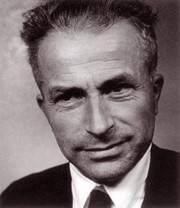
Albert Frey-Wyssling

Laureate 1949: Albert Frey-Wyssling

“[…] for his work on submicroscopic morphology published in 1949 […]. Following many years of systematic work researching the fine structure of cell walls and plant plasma components that are invisible under an optical microscope, he observed that submiscroscopic morphology offered the same perfect forms and complicated problems of form as the optical miscroscopic universe. It was therefore hoped that the same method could be used in future to reveal the fine structure of animal and human cells.”

Die einzigartige Leistung F.s bestehe laut den Gutachtern nicht zuletzt in der Tatsache, dass er den von ihm zuerst nur indirekt erschlossenen Feinbau der Zellbestandteile anschliessend mittels elektronenmikroskopischer Untersuchungen bestätigen konnte. Auf diese Weise sind ihm Abbildungen von verschiedenen seiner Voraussagen gelungen, so vom Schichtenaufbau der Chloroplasten, von der Mikrofibrillenstruktur der Zellwände sowie vom Gelbau von Bakterienzellulose (Retikularstruktur).

Contact

Marcel Benoist Foundation
State Secretariat for Education,
Research and Innovation SERI
Einsteinstrasse 2
CH – 3003 Bern

  • Legal notice