• de
  • fr
  • it
  • en
  • Laureates
    • Laureate 2024
    • Prize 2024
    • Past laureates
    • Subsequent Nobel Prize laureates
  • Nomination
    • Nomination 2025
  • The award ceremony
    • The award ceremony 2024
    • The award ceremony 2023
    • The award ceremony 2022
    • The award ceremony 2021
    • The award ceremony 2020
    • The award ceremony 2019
    • The award ceremony 2018
    • The award ceremonies of the past
  • Activities
    • Lecture 2025
    • Workshop 2024
    • Lecture 2024
  • Marcel Benoist
    • The founder
    • The legacy
  • Foundation
    • Foundation
    • News
    • Board of Trustees
    • Patronage Committee and Friends
    • Create excellence!
    • Foundation assets
    • Contact

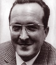
Hans J. Moor

Laureate 1967: Hans J. Moor

“[…] for their important work on the development of the freeze-etching procedure as a method of preparing plant, animal and human cells. Their discovery allowed the ultrastructure of cells to be realistically represented in the high vacuum of an electron microscope and earned them the highest respect of the scientific community.”

Das Gefrierätzverfahren besteht darin, zuerst die kurz auf -100°C eingefrorenen Zellen anzuschneiden, das Eis im Hochvakuum wegzusublimieren (‘Ätzung’) und vom entstandenen Relief durch Schrägaufdampfen von Platin einen Abdruck zu erstellen, der dann im Durchstrahlmikroskop ein negatives Oberflächenbild ergibt. Diese ursprünglich zur Untersuchung von Viren angewandte Methode sei von den beiden Preisträgern in die zytologische Ultrastrukturforschung eingeführt worden. Mit ihrer Pionierarbeit, so die Experten, eröffneten sich grosse Perspektiven in der Physiologie und der Krebsforschung.

Contact

Marcel Benoist Foundation
State Secretariat for Education,
Research and Innovation SERI
Einsteinstrasse 2
CH – 3003 Bern

  • Legal notice